Day 1 :
Keynote Forum
Ana Falcon
National Center for Biotechnology, Spain
Keynote: Severe outcome in influenza virus infected patients is associated with reduced accumulation of defective viral genomes
Time : 10:25-11:00

Biography:
Ana Falcón completed her PhD in Molecular Biology and Science at Autonomous University, Madrid. During her scientific career, she has studied molecular biology of several respiratory viruses of clinical significance as influenza virus, SARS-CoV and other coronaviruses. She has worked on virus reverse genetics, functional analysis of viral and cellular proteins, and molecular diagnosis of virus in clinical samples and on animal models to study viral pathogenesis. She is developing and coordinating an international multidisciplinary project including physicians, microbiologists, molecular virologists and bioinformatics. She is a Reviewer for several scientific journals and has trained PhD and master students.
Abstract:
Influenza A virus (IAV) infection can be severe or even lethal in toddlers, the elderly and patients with certain medical conditions. Infection of apparently healthy individuals nonetheless accounts for many severe disease cases and deaths, suggesting that viruses with increased pathogenicity co-circulate with pandemic or epidemic viruses. Looking for potential virulence factors, we have identified a viral genetic determinant that contributes to infection outcome. A polymerase mutation identified in a fatal IAV case, when introduced into two different recombinant virus backbones, led to reduced defective viral genomes (DGs) production and increased pathogenesis in mice. These data provide genetic support for the association of pathogenicity and low DGs accumulation induced by mutations present in pathogenic viruses circulating in humans. Testing this association, we performed a genomic analysis of viruses isolated from a cohort of previously healthy individuals who suffered highly severe IAV infection requiring admission to intensive care unit, and patients with fatal outcome who additionally showed underlying medical conditions. These viruses were compared with those isolated from a cohort of mild IAV patients. Viruses from highly severe/fatal outcome patients showed significantly fewer DGs accumulation than control viruses, suggesting that low DGs abundance constitutes a new virulence viral pathogenic marker in humans, regardless of the mutations responsible.
- Viral Infections | Bacterial diseases | Antimicrobial Agents and Antimicrobial Activity | Fungal Diseases | Pharmaceutical Microbiology | Engineering Biotehnology
Location: Waterfront
Chair
Godfred Menezes
Hail University, UAE
Session Introduction
Vasundhara Rangaswamy
Stanford Hospital and Clinics, USA
Title: Comparing diagnostic microbiology in 2 worlds
Time : 11:20-11:45

Biography:
Vasundhara Rangaswamy completed her MD from India and MS Clinical and Molecular Biology from California. She currently works in the clinical microbiology lab of Stanford Hospital and Clinics, California. She has been involved in lab capacity building in India, Cambodia and Ethiopia and also in public health initiatives in rural India for last 20 years. She has also been involved in teaching clinical microbiology and rural health issues to all levels of health care professionals.
Abstract:
Aim: Microbiology is the pillar of infectious disease diagnosis and treatment. Strengthening of services globally is mandatory to achieve health goals laid out by many organizations. However, diagnostic microbiology services offered even for diseases like Malaria, HIV, TB or diarrhoeal diseases that are prevalent in poor countries, differ remarkably between various labs. The presentation attempts to compare an advanced microbiology lab in California, with labs Cambodia, Africa and rural India.
Methodology & Observations: The observations are based on personal experiences of working in or visiting labs in different countries and settings. For any lab, compared to routine tests offered in haematology, biochemistry, serology, and urinalysis, providing reliable basic microbiology services is challenging. In less developed regions, it is a herculean task. Some of the many challenges faced are, lack of awareness among physicians and common folk about the contribution of clinical microbiology towards patient care, absence of or little regulation on QA/QC systems, formidable costs of infrastructure and unreliable supply of water or electricity. One is confronted with many burning questions. Is the disparity in different settings fair? Are the goals same? Whose responsibility is it to step up lab capacity building? Despite these obstacles, significant changes are taking place in some health care centers due to the efforts of one odd passionate microbiologist, pursuance by astute physicians, remarkable work of organizations like LabCAP, DMDP, CDC, etc., by inventors who have simplified diagnostic tools and made them affordable yet reliable and by global pressure to step up containment of diseases.
Conclusion: More of us need to get involved; efforts need to be constant and probably faster to keep pace with the growing population and spread of diseases.
Godfred Menezes
Hail University, UAE
Title: Evaluation of synergistic activity of lactoferrin with antibiotics against drug resistant bacterial pathogens
Time : 11:45-12:10

Biography:
Godfred A Menezes is currently working at RAK College of Medical Sciences (RAKCOMS), RAKMHSU, UAE. He was an Assistant Professor and Scientist at Hail University, Saudi Arabia; In-charge/Scientist of Central Research Laboratory (CRL) and; Assistant Professor of Microbiology at Sree Balaji Medical College & Hospital, Chennai, India for three years. He has also worked as a Scientist in the Department of Medical Microbiology and Infectious Diseases, Netherlands. He has been worked extensively on molecular characterization of antimicrobial resistance in clinical bacterial pathogens. He has been a faculty of several medical institutes and also a Para-Medical Institute.
Abstract:
The multidrug-resistant (MDR) bacterial infections have escalated as one of the world's utmost health issues. The progress of novel antibiotics has declined over the last half century. The aim of this study was to determine the effect of lactoferrin (human, bovine and camel) on minimum inhibitory concentrations (MICs) of important antibiotics in use against drug resistant bacterial pathogens encountered in the region of Hail, Kingdom of Saudi Arabia (KSA). Totally 147 clinical bacterial isolates were successfully isolated. Pathogens included were: Methicillin resistant Staphylococcus aureus (MRSA)-30 isolates; methicillin resistant coagulase negative Staphylococcus-30 isolates; extended-spectrum beta-lactamase (ESBL) producing Enterobacteriaceae-40 isolates; fluoroquinolone resistant gram negative pathogens-30 isolates; multidrug resistant Pseudomonas species- 05 isolates; carbapenem resistant gram negative pathogens- 05 isolates; AmpC beta-lactamase producing gram negative pathogens- 05 isolates; vancomycin resistant enterococci (VRE)- 02 isolates. The methods employed were MALDI-TOF for identification, MicroScan WalkAway system for identification, susceptibility testing and LF synergism testing. PCR-Sanger sequencing was done (before and after exposure to LF synergism) to study the molecular biology aspects of the study. In our study, the reproducible synergism effect of LF with antibiotics was found to be remarkable. To specify the phenotypic effects of LF in synergism with antibiotics: the isolates producing ESBL had turned non-ESBL; quinolone resistant isolates had turned susceptible; MRSA had turned MSSA (methicillin susceptible) and VRE had turned susceptible. The molecular biological study suggests changes only in the gene expression after the exposure to LF compounds. The results of this study demonstrated similar effect with comparable results for the LF tested from three different sources (human, bovine and camel). The outcome knowledge of the study would certainly help the Ministry of Health (MOH) in planning the LF adjuvant treatment methods for locally faced drug resistant pathogens causing different infections.
Anne Elain
Université de Bretagne-Sud, France
Title: A fluorescence-based method for the assessment of polyhydroxyalkanoates (PHA) production
Time : 12:10-12:40

Biography:
Anne Elain received her PhD degree in Process Engineering from Rennes I University, France, in 1999. She then joined the Biomaterials and Nanotechnologies Lab (now IRDL) of Bretagne Sud University, France. Currently, she leads the Department of Biochemical Engineering. Her general research interest areas are applied microbiology and fermentation technology for the production of high value-added products and process optimizing (yield, sustainability, economic cost, etc.).
Abstract:
Polyhydroxyalkanoates (PHAs) are polyesters acting as energy and nutrition reserves for many prokaryotes. The substrate, cultivation strategy and production strain, will determine the chemical nature and the physical characteristics (i.e., molecular weight, tensile strength, melting temperature, degradability timelines, etc.) of these polyesters. This opens the door for substituting petrol-based thermoplasts, elastomers or latexes, in multiple industrial and biomedical applications. For instance, in the biomedical field, the great adaptability of PHA, in association with their biocompatibility and their bio-absorption capabilities, make them attractive for use as in vivo implants, regeneration devices or drug delivery systems (3, 4). Until now, the major challenge is to reduce the cost of the biosynthesis process to permit the development of a reliable and sustainable PHA production chain. One aspect of major economic importance is to carefully optimize the operating conditions in order to maximize biomass growth and polymer yield. Hence, a rapid and reliable method of screening and monitoring process performances (i.e., cell growth and PHA contents), is needed. Although, GC-MS analysis provides the most accurate results relative to PHA quantification and monomer composition, it involves extraction and derivatization steps which are complex and time-consuming when applied to a large number of samples. In this study, we developed a method that used the lipophilic fluorescent probe Nile Red (1 mg L−1 solution in DMSO) directly into the culture broth to stain the PHA inclusions inside bacterial cells followed by detection of the emitted fluorescence by both microscopic and spectrometric techniques. Epifluorescence microscopy provides a rapid tool to distinguish producing from non-producing bacterial species and the relative fluorescence intensity (FI) determined at the maximum of emission spectra in the wavelength region of 560-710 nm, allows a fast assessment of the cultivation condition and physical process that may enhance PHA production yield. The method was found effective to select bacterial strains efficient for PHA synthesis among a marine collection. Subsequently, the NR assay was used to determine the C0/N0 ratio of the producing media that may improve the polymer yield as well as to follow the time course of fermentation. The coupling of fluorescent dye staining to epifluorescence microscopy is thought to open up new possibilities for high-throughput screening applications and identification of novel PHA producers.
Ka Tik Cheung
Tung Wah College , Hong Kong
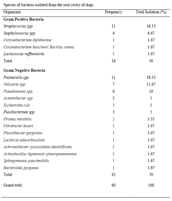
Title: Evaluation of domestic dogs and cats oral flora in relationship of oral hygiene and detection of multi resistant bacterial strains
Time : 12:40-13:10

Biography:
Ka Tik Cheung is a Lecturer at Tung Wah College since 2013. He has completed his Doctoral degree in Microbiology and has several years of experience in clinical laboratory. He has also acquired the license to practice which is certified by The Hong Kong Medical Laboratory Board. His research focuses on “Veterinary infectious disease and antibiotics susceptibility on companion animals”. He is also actively involved in zoonotic disease research. He is a member of Hong Kong Veterinary Nursing Association.
Abstract:
Recently, human-animal interaction has occurred more frequently, animal bite injuries have become a serious and high-risk problems as the result. Oral flora can be transferred by close oral contact and through bites. While most of the bites do not require medical progress, only some bites would become an infection. The resultant infection is typically a poly-microbial infection; consist of common environmental flora and the oral flora in animals. As oral hygiene is an important method to reduce the number of bacteria of human oral cavities, but there are only a few articles demonstrated in domestic dogs and cats. To evaluate the effectiveness of oral hygiene in domestic dogs and cats, this study compared the complexity of isolates from the oral cavities and the frequencies of performing oral hygiene. The age of domestic dogs and cats was compared to the number of isolates from the flora as well. Besides, the oral flora was identified and the frequency of occurrence was evaluated, in order to attempt the bacteriology of domestic dogs and cat’s oral cavity and the causative agent of human infection. In this study, gram stain, a series of biochemical tests and Matrix-assisted Laser Desorption Ionization-Time of Flight Mass Spectrometry (MALDI-TOF-MS) were used in identification. Pasteurella species and Streptococcus species were isolated in high frequency. Neisseria species, Pseudomonas species, Enterobacteriaceae family, Corynebacterium diphtheriae, Achromobacter xylosoxidans/denitrificans and Sphingomonas paucimobilis were also identified in the samples as well. Moreover, detection of multiple antibiotic resistant bacteria was also carried out in this study as well. It was used to provide more evidence-based information on antibiotic therapy in dog bite wounds and related infections. In which, multi-resistant organisms and extended-spectrum beta-lactamase (ESBL) positive Enterobacteriaceae were found in the oral cavity of sample dogs. Antibiotic susceptibility patterns for some bacteria were evaluated in this study.
Debabrata Biswas
National University of Singapore, Singapore
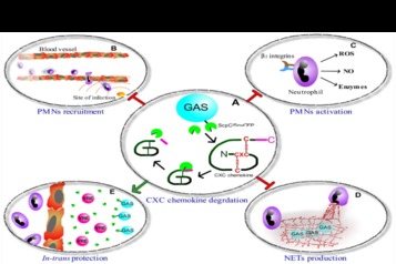
Title: Group A Streptococcal CXC chemokine-protease cleaves the human antimicrobial cathelicidin peptide LL-37 resulting in the loss of immunomodulatory functions of the peptide
Time : 14:10-14:35

Biography:
Debabrata Biswas completed his PhD in the field of Cell Biology while working on Arsenic Toxicity in blood. He shifted his interest to the study of pathogen biology and infectious disease during his Post-doctoral study at Hebrew University of Jerusalem. He joined the laboratory of Prof Emanuel Hanski, working on the mechanism of pathophysiology of group A streptococcal soft tissue infection. His present work in Microbiology department of National University of Singapore deals with bacterial virulence factors and various strategies that might be designed against the disease based on the mechanism of actions of these factors. He is currently investigating molecules in the host inflammatory and immune signaling cascade that might act as the potential targets of the bacterial serine protease ScpC, which is a major virulence factor in soft tissue infections.
Abstract:
Statement of the Problem: Severe-soft tissue infections caused by group A streptococci (GAS) are characterized by a rapid dissemination of GAS followed by massive necrosis and tissue destruction. The human antimicrobial peptide, LL-37, is expressed during invasive GAS infections. It is believed that LL-37 antibacterial activity limits GAS spreading, since mice deficient of the LL-37 murine analog, CRAMP, is more sensitive than wild type mice to subcutaneous GAS challenge. LL-37 also directly recruits neutrophils to the site of infection and stimulates interleukin-8 (IL-8) production by keratinocytes. Thus, the immunomodulatory activity of LL-37 is aimed to exacerbate neutrophil response that is crucial for eradication of GAS from soft-tissue. Yet, analysis of debrided human soft-tissue samples revealed the coincidence of LL-37 along with viable GAS.
Theoretical Orientation: The GAS CXC-chemokine protease ScpC plays a central role in virulence through IL-8 cleavage preventing recruitment of neutrophils to the site of infection and reducing the production of neutrophil extracellular trap. Because of its immunomodulatory activities, we hypothesized that LL-37 may also serve as a substrate for ScpC. Functional significance of ScpC-mediated cleavage of LL-37 was studied in vitro and verified in vivo in the mouse model of human GAS soft-tissue infections.
Findings: Here, we demonstrate that immunomodulatory activity of LL-37 is crucial for controlling GAS spreading in soft-tissue. We found that GAS CXC-chemokine protease ScpC degrades and inactivates IL-8 as well as LL-37. ScpC cleaves the first 8 amino acids from the N-terminal of LL-37. This results in loss of its capacity to recruit neutrophil and stimulate IL-8 production by keratinocytes. In summary, the capacity of ScpC to shut down recruitment of neutrophils that is mediated through both IL-8 and LL-37 as well as inactivate LL-37-mediated IL-8 production by keratinocytes reflects a perfect adaptation of GAS to its human host.
Urszula Kosikowska
Medical University of Lublin, Poland
Title: Activity of novel N-substituted-pyrazoles, dichlorobenzoylthiosemicarbazides, 2,4-thiazolidindiones and 1,2,4-triazole-ciprofloxacin hybrids on Haemophilus parainfluenzae and Haemophilus influenzae planktonic and biofilm-forming cells
Time : 14:35-15:00

Biography:
Urszula Kosikowska (PhD) is a Pharmacist, specialist in Medical Microbiology. She is a Lecturer in the Department of Pharmaceutical Microbiology with Laboratory for Microbiological Diagnostics, Medical University of Lublin, Poland. She has published scientific publications in reputed journals, popular articles and conference reports. Her research interests include issues of respiratory microbiota, diagnostics and drug resistance of bacteria and antimicrobial activity of newly synthesized compounds on planktonic and biofilm-forming cells mainly of the genus Haemophilus and other selected members of the family Pasteurellaceae.
Abstract:
Biofilm is a single- or multi-species highly specialized and multi-functional population of microorganisms living on natural or synthetic surfaces with specific biological and physicochemical properties. Biofilm is also an important factor in the pathogenesis of e.g. opportunistic infections. Among the important features of the biofilm cells is their resistance to drugs and other antimicrobial agents and host defense factors. Consequently, this may lead to therapeutic failures. Hemophilia, mainly Haemophilus parainfluenzae and nontypeable Haemophilus influenzae (NTHi), are opportunistic representatives of the respiratory microbiota which are able to live as biofilm and to cause recurrent or chronic and systemic infections (e.g. endocarditis, bronchitis and bacteremia or meningitis). On the basis of minimal inhibitory (MIC) and minimal biofilm inhibitory (MBIC) concentration values the differences in the effect of newly synthesized compounds on planktonic and biofilm-forming cells of hemophilia was observed. Among all the tested derivatives the highest activity against both planktonic and biofilm-forming cells of H. parainfluenzae was estimated for 1,2,4-triazole-ciprofloxacin hybrids and for N-ethyl-3-amino-5-oxo-4-phenyl-2,5-dihydro-1H-pyrazole-1-carbothioamide against both planktonic and biofilm-forming cells of H. parainfluenzae (including ampicillin-resistant strains) and H. influenzae. The basis for the activity of these compounds was the inhibition of enzymes important for DNA synthesis and cells’ metabolism as well as anti-adhesive properties in the early stages of biofilm formation. Other compounds, e.g. thiosemicarbazides and thiazolidinediones did not have a substantial inhibitory effect on both planktonic and biofilm-forming cells. The effect of the tested derivatives partially depends on species or strain of bacteria, compound’s structure and concentration, and time of incubation.
Tseaa Shambe
Benue State University, Nigeria
Title: Postharvest losses of yam tubers in Benue state, Nigeria, West Africa
Time : 15:00-15:25

Biography:
Tseaa Shambe is a Chemist and a Lecturer in Organic Chemistry. He has published papers on chemical composition and structures of some carbohydrates and their degradation by enzymes and acids. He has also worked on the use of bread and composite flour for bread and confectionaries. He is also interested in food toxicology and works very closely with microbiologist.
Abstract:
Statement of the Problem: Africa is the largest producer of yam in the world, with the highest production coming from West Africa. Benue State is the largest producer of yams in Nigeria followed by other States in North Central region. This is because of the rainfall, soil and other climatic conditions that are favorable for yam production in this region. The yams produced form staple food for about 182.2 million people. Postharvest losses of yam tubers (Dioscorea rotundata and D. alata) may occur from a number of causes ranging from improper handling of the tubers to bio-deterioration by microorganisms, insects or rodents. The largest cause of postharvest losses of yam tubers is from microorganisms.
Methodology & Theoretical Orientation: Isolation and identification of microorganisms responsible for the rot of yam tubers was carried out using standard methods of isolation and identification. Optimum temperature of growth was analyzed; pathogenicity test was conducted on the isolates to confirm them as the etiologies of the rot. Plant extract was prepared and incorporated on media plates and used for antimicrobial sensitivity test.
Findings: Four bacteria species (Serratia marcescens, Erwinia carotovora, Klebsiella oxytoca and Pseudomonas aeruginosa) and five fungi species (Aspergillus niger, Rhizopus stolonifer, Botryodiplodia theobromae, Fusarium oxysporum and Penicillium marneffei) were consistently isolated in samples from the various sampling areas. Pathogenicity test revealed the organisms as the cause of the rot. They were inhibited by the plant extracts partially or completely.
Conclusion & Significance: Extensive loss of the harvest can be prevented by blending the various extracts and spraying them on the yams to arrest rot.
Leonardo Madrucciani
Advisor Petroleum LTD, UK
Title: Viral Detection by Nano-amplification Micro Array Technique
Time : 15:25-15:50

Biography:
Abstract:
Statement of the Problem: Microarray as a novel biological technique has been used in gene detection widely. But this method needs improvement, considering expensive probe labeling material and valuable signal scanner. So a visual detection technique was established by applying sandwich hybridization and silver stain. This technique was 100 times as sensitive as LCS (laser confocal scanning) and made it possible to develop a rapid and cheap detection method for Virus. Increasing studies revealed the application of gold nanoparticle as oligonucleotide label was wide.
Methodology & Theoretical Orientation
Materials of Viral detection microarray
Preparation of gold nano-particles described by Grabar (Grabar et al., 1995)
Preparation of detection microarray for Virus
Findings: Cumulative evidences showed gold labeled probe was efficient and could be alternative marker in microarray assay. On the other hand, the sliver stain enhancement, a technique companied with nano-gold labeling oligonucleotide, was used to amplify detection signal of nano-gold. This technique made detection signal visual and its sensitivity of gene detection was approximately 1000-fold high as that of Cy3-based fluorescence.
Muhammad Riaz
University of Agriculture, Pakistan
Title: PCR-RFLP, a diagnostic tool for rapid detection of drug resistant Mycobacterium tuberculosis
Time : 16:10-16:35

Biography:
Muhammad Riaz has PhD degree in Biochemistry from University of Agriculture, Faisalabad, Pakistan and completed his six months research training at University of Glasgow, Scotland, UK. He is working as a Lecturer in the Department of Allied Health Sciences, Sargodha Medical College, University of Sargodha, Pakistan. He has published more than 15 papers in reputed journals. He participated and presented research papers in international conferences as oral and poster presentation.
Abstract:
Tuberculosis (TB) is a highly contagious disease caused by Mycobacterium tuberculosis. Roughly 1/3rd of the world’s population is infected with Mycobacterium tuberculosis (MTB). So, the current study was carried out to determine the drug resistant Mycobacterium tuberculosis magnitude through PCR-RFLP. Initially, the patients were screened for tuberculosis through sputum smear microscopy by Zeihl Neelsen (ZN) staining technique. The sputum positive patients were included in the study after informed consent to the patient. A total of 341 patients were included in the study. PCR-RFLP was used to evaluate the variation in genetic makeup of drug resistant mycobacterium tuberculosis strains. Among the studied population, individuals in older age are affected more and also TB is more common in uneducated and poor people. Resistance against isoniazid, streptomycin, ethambutol and ofloxacin were studied. Overall 91.5% patients were confirmed positive for M. tuberculosis complex infection on PCR analysis. It was found that 17.30% of the cases in the study population have multidrug resistant tuberculosis (MDR-TB). Co-infection of TB with diabetes, HCV and HIV were also observed. Among the drug resistant TB cases 24.04% of the cases were resistant to isonizid (INH), 19.87% resistant to ethambutol (EMB) and 16.99% resistant to streptomycin (STRM). Fluoroquinolone namely ofloxacin resistance along with ethambutol, isoniazid and streptomycin was observed in 6.5% of cases. We concluded PCR-RFLP as a useful molecular technique for rapidly detecting mutations in drug resistant TB patients.
Shimuye Kalayu Yirga
Woldia University, Ethiopia
Title: Preparation and characterization of monoclonal antibodies against citrinin (CTN) for immunoassay VP
Time : 16:35-16:55

Biography:
Shimuye Kalayu Yirga has done his PhD student in School of Life Sciences, Fujian Agriculture and Forestry University in China. Since 2013, he has been working as a Lecturer at Woldia University in Ethiopia. His PhD research focuses on molecular immunology, production of monoclonal antibody and antibody cancer therapy. Recently he found an IgG2a, IgG2b and IgG3 monoclonal antibody against kidney toxin and characterized using different immunoassay. He has done his Bachelor’s degree from Haramaya University, Master’s degree from Bahir Dar University in Ethiopia.
Abstract:
Citrinin (CTN) is a hepato-nephrotoxic mycotoxin produced by fungi genera of Aspergillus, Monauscus and Penicillium. CTN contaminates grains, fruits, juices and vegetables cause various toxic effects to humans and animals. The commonly used analytical methods for CTN detection were TLC), HPLC, LC-MS/MS, (GC-MS). However, these methods have lack of potential for automation, low sensitivity, low specificity, complex equipment’s, incompatible with the real sample, energy and time consuming. Citrinin is a small non-immunogenic toxin with a molecular weight of 250.25 g/mol. Thus, it is necessary to conjugate it with carrier proteins for an immuno-response to generate antibodies. Different methods were applied to conjugate CTN with carrier proteins. Bio coupling method was not satisfactory due to difficulties in removing unconjugated materials through dialysis furthermore; in our study the formaldehyde condensation method was also not effective with the conjugation of CTN-OVA because the result of conjugation was not stable. Finally , in this study CTN was conjugated to bovine serum albumin (BSA) and ovalbumin (OVA), respectively, by amide bonds through active ester method using 1-ethyl-3-(3-dimethylaminopropyl) carbodiimide hydrochloride (EDC) and N-hydroxysuccinimide (NHS). In this study, Female Balb C mice were immunized with CTN-BSA conjugate and Obtained a high anti-serum titer (1:32,000 v:v) when compared to the non-immunized control mouse. Spleen cells of the immunized mice were fused with Sp2/0 myeloma cells to obtain 21H27 hybridoma cell. Ascitic fluid of hybridoma cell was produced in mice abdomen, and purified using caprylic/ammonium sulfate precipitation method. The 21H27 anti-CTN mcAb was the IgG2a antibody subclass, and cross-reactivity results indicated that anti-CTN mcAb is specific to CTN with high affinity (2.0-108 L/mol). Indirect competitive ELISA (IC-ELISA) results showed that the linear range of detection was 0.01–5.96 ng/mL and the IC50 was 0.28 ng/mL with a lower detection limit (LOD) of 0.01 ng/mL. The average recovery was 93.8%-1.6% with a coefficient variation of 1.0%-4.3%. Hence, anti-CTN mcAb secreted by 21H27 hybridoma cell was successfully produced and can be used to detect CTN contaminated feed and foodstuffs.
In this study, 2 mL of sodium citrate and 8.2 pH adjusted with potassium chromate was applied for preparation of good color gold solution. In addition to this, freshly prepared (within one week shelf life) anti-CTN mcAb gold conjugated and optimum concentration of coating antigen were the appropriate parameters for the best achievement of anti-CTN mcAb immuno-chromatographic test strip assay.

Figure 1 Graphic summary of monoclonal antibody production against Citrinin for immunoassay.